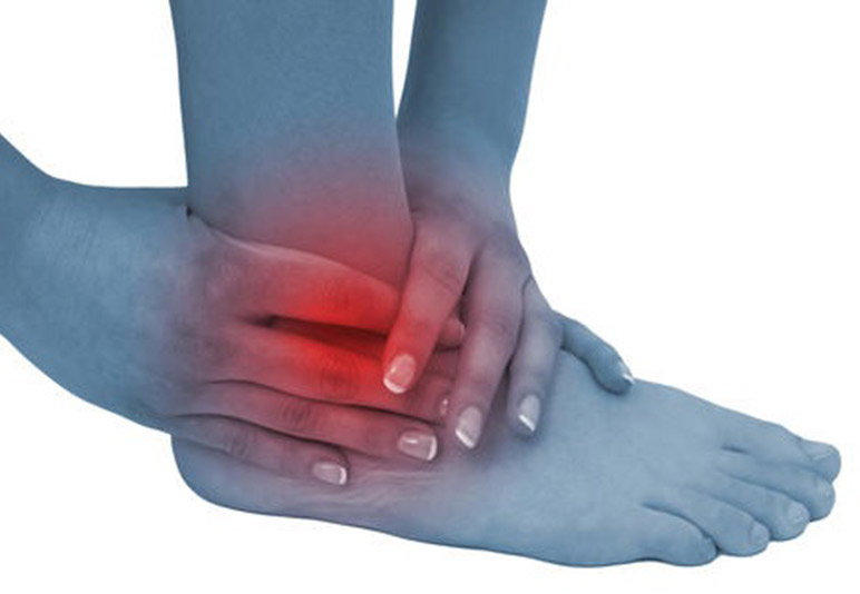
Persistent ankle pain can limit activity and disrupt the ability to comfortably carry out the most simple activities of daily living. The severity of symptoms can vary from individual to individual. Causes for generalized ankle pain and impingement can include 1) joint damage, 2) damage to the ankle joint capsule and 3) ligament and tendon damage.
These painful conditions can be acutely initiated following an injury as basic as twisting an ankle. Other factors that can contribute to bone, soft tissue and joint damage include generalized repetitive motions over a long period of time which can weaken anatomic structures resulting in overuse syndromes. In some cases, these conditions can be treated conservatively with a combination of injections, physical therapy, bracing/orthotics and anti inflammatory medications. Surgery, however, is often indicated and can prove very effective in treating certain ankle conditions.
Ankle pathology such as torn ligaments, joint cartilage damage, ankle joint capsule synovitis and loose floating pieces of cartilage/bone can all be treated via arthroscopic surgery. These conditions, typically identified through physical examination and MRI, can cause ankle joint impingement, limitation of ankle joint motion as well as joint pain with activity. In addition, many patients note clicking and popping sounds audibly recognized as emanating from the joint itself.
Surgical techniques typically fall into one of two major categories – traditional open procedures vs. minimally invasive techniques. Ankle arthroscopy is a surgical procedure which is minimally invasive and provides the surgeon access to the interior of the joint in order to repair physical damage to the affected structures. The technique is traditionally performed via two very small incisions at the anterior aspect of the ankle which allows for the introduction of a small camera or ‘scope’ and specialized instruments into the joint space. The live arthroscopic images are viewed during surgery on a television monitor in real time.
Through this arthroscopic technique regions of cartilage damage can be removed and micro fractured to encourage new tissue growth. Torn ligaments can also be debrided and repaired. Synovitis, as well, can be resected along with the removal of bone spurs and loose floating pieces of bone and cartilage found in the joint.
Post operative recovery can range from 7 – 21 days depending upon the underlying pathology addressed. In many cases physical therapy and active range of motion exercises are prescribed. In some instances patients are encouraged to begin waking within 24 hours of surgery. The limited incision size reduces recovery and provides the added benefit of limited scar tissue formation in comparison to more traditional surgical approaches. The majority of patients report an improvement in joint function and mobility following the procedure.